An Expert Guide to Mole Removal

Vicky Eldridge
By Vicky Eldridge

Vicky Eldridge is an award-winning journalist, editor and copywriter, with 18 years’ experience in aesthetics.


The NHS no longer delivers “non-essential” mole treatments, meaning that the majority of mole removal procedures, or the removal of skin ‘lumps and bumps’ (e.g., warts, verrucae, and skin tags), can no longer be administered for free on the NHS. This means that private medical practices and aesthetic clinics are increasingly seeing patients come through the doors wanting procedures like mole removal. 
 
Mole removal is a very popular skin treatment. It can be life-saving, but it deserves a bit more contemplation. Whether you are offering mole removal at your clinic or referring patients elsewhere, it’s important to be educated about moles. Early detection is key, and aesthetic clinicians may be the first to notice changes in the skin if a patient comes in regularly for treatment and has any visible moles. 
1. “Mole removal”
There is already a diagnosis in this term: mole. What is a mole? A mole or nevus has a melanocytic origin, just like the dangerous melanoma skin cancer. So, when you think about ‘mole removal,’ make sure that the diagnosis is right. Whoever performs the mole removal should be able to diagnose it correctly first, which is not always easy or straightforward. I have seen cases when a melanoma was removed with an ablative laser and without histological verification. The next things the patient noticed were the metastases or the spreading tumour.
 
Conclusion: Mole removal should be performed or initially checked by a dermatologist first in a skin consultation.
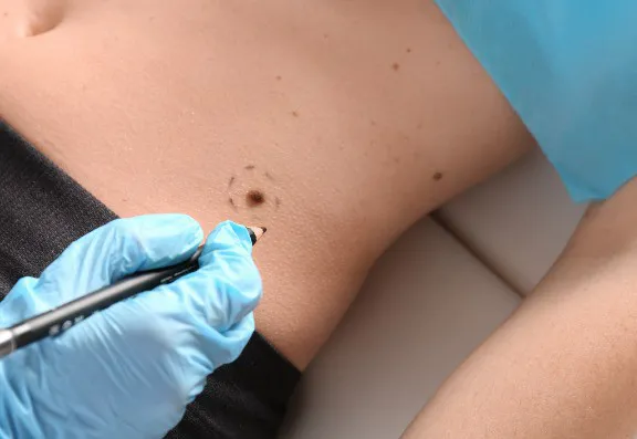
Mole removal
2. Reason for mole removal
There are multiple possible reasons for mole removal: an unsightly mole, an annoying lesion, frequent irritation, or a suspicion of a malignant transformation. These reasons weigh differently when we make pro-con mole removal decisions. For example, a small blemish on the chest that you don’t like is not a good enough reason to risk a keloid, which is overgrowing and disturbing scar tissue. A suspicious lesion at the same location has to be removed without hesitation. The chest is a high-risk area for abnormal healing (keloid formation).
 
3. Location of the Mole:
Different parts of the body have different healing potentials. Generally, the face heals well and quickly, but it has a very high cosmetic significance. The worst healing potential on the body is the triangular area on the chest, with the highest chance of keloid formation. The back is an area where the skin is very thick due to the strong forces on the skin every time we bend forward, and it has to be considered when making decisions.
 
4. Photography before skin treatments
Photography before any skin procedure is of the utmost importance. Taking pictures has a medico-legal requirement and shows that you care and are willing to learn from your work. Taking pictures may help you notice any changes to the patient’s skin (and any moles) between treatments. They are also a valuable way of showcasing results to other patients with appropriate consent. 
 
Mole removal method
5. Mole removal method
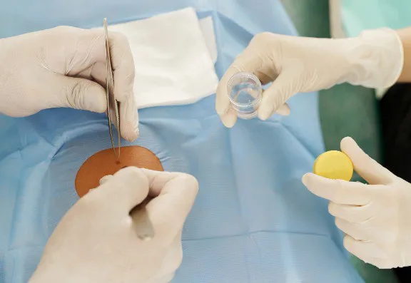
There are different methods for mole removal. The most suitable one should be selected based on different factors, such as the nature of the lesion, whether it is flat or elevated, the location, histology needed or not, etc. If the lesion is a mole and it is flat and looks asymmetrical, the method to use is excisional biopsy with a narrow safety margin followed by histology analysis. Shave biopsy can also be used to remove suspicious lesions. Ablative removal methods that destroy the lesion entirely, such as laser, cryotherapy, and electric cauterisation, should be used only carefully in expert hands who are experienced enough to distinguish a benign lesion from a potentially malignant one.
6. Safety margin
Specific lesions require a safety margin when removed. When we remove moles, we typically leave a small free margin around the lesion, so there is nothing left back with the potential to regrow. When inadequately removed, moles may grow back, and they can look ugly and develop into pseudo-melanoma, which causes some headaches for clinicians and pathologists alike. With an ablative method (laser, cryo, cauterisation), the extent of the safety margin is unclear.
 
7. Histology analysis after mole removal
In every case, histology analysis should be performed when there is the slightest suspicion about a skin lesion being malignant or the lesion’s nature is unclear. It will reveal the final diagnosis, and it can determine whether any further action is needed or not.
 
8. The scar after mole removal
There is a very simple rule here: any procedure that reaches the dermis (the second, thick layer of the skin) causes a scar. Procedures more superficial to this don’t leave a scar. So, small lesions such as viral warts, fibromas, and seborrheic keratoses can be removed without causing a permanent scar. If completely removed, any lesion extending into the dermis (most of the moles) leaves a permanent scar. Moles can be removed superficially with a shave biopsy, but in this case, it is most likely that there are remaining deeper cells with the potential to reappear.
 
9. Risks and complications
During the consultation process, patients need to understand clearly the potential risks and complications of mole removal, such as infection, recurrence, unsatisfactory healing (keloid, hypertrophy, pigmentation), or unintended ablative removal of a malignant tumour without histology, including the possibility when no procedure is done.
 
10. Aftercare
It depends on the location and size of the scar, but generally, a fresh scar needs support and protection. The dressing should be kept on for a few days without getting wet. Patients should avoid physical activity at this stage. After the removal of stitches (if applicable), a firm massage with cream or oil is recommended multiple times a day (the more, the better), followed by the application of a silicone liquid or tape. Scars need UV protection for 18 months post-procedure.
 
Conclusion
Mole removal, while common, is not a decision to be taken lightly. It involves considerations regarding diagnosis, location, method, and aftercare, each with its own complexities. To ensure the best outcomes, it is crucial patients consult with a skilled dermatologist who can guide them through the process with expertise and care.
 
This article was written for the Consulting Room Magazine.
 
Fancy grabbing a FREE subscription to the Consulting Room quarterly aesthetic trade magazine?
Immerse yourself in the world of aesthetic medicine with our captivating Consulting Room Magazines. Each edition is carefully curated with CPD-certified content, delving into various sectors or topics within the field. Our unique quarterly themed editions take a deep dive into different sectors or topics associated with the world of aesthetic medicine with articles written by leading practitioners in their fields of expertise.
 
Get your free subscription now at: https://www.consultingroom.com/magazine/

Unlock a treasure trove of aesthetic knowledge!

To access a digital archive of all previous magazines and articles, including this one, simply provide your details here and enjoy lifetime access to our digital magazine archive. Explore past issues and delve into a wealth of valuable content.
Rec

Keep In Touch

Ensure you and your staff stay up-to-date with key topics shaping the field of aesthetics.

Your free digital round-up of relevant aesthetic news articles and trending items delivered directly to your inbox.

Immerse yourself in our quarterly, complimentary, themed digital magazine, compiled by award-winning editor Vicky Eldridge.

Stay informed of new technologies and receive exclusive news and offers from carefully selected aesthetic partners.